Bio-Medical Imaging
within the activities of the
CDS-group
(formerly
CDS-Lab), ICAR-CNR
Objectives
The activity concerns the analysis, design, and implementation of machine learning methods for multimedia applications involving bio-medical images and image sequences.
Main achievements
Dermoscopic images
 |
Approach to the characterization of uncertain lesions, to
detect characteristic profiles of benign and malignant lesions [2].
Based on suitable multi-value descriptors (scalar, interval, and histogram data) extracted by dermoscopic images, it consists in
selecting through discriminant analysis the most discriminating features and detecting through dynamic clustering the characteristic profiles.
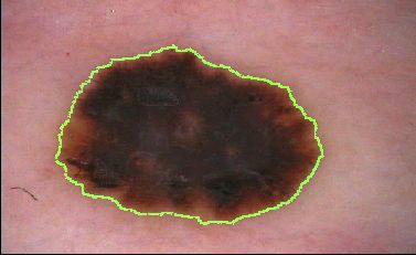
For the automatic segmentation of skin lesions in dermoscopic images, we proposed the SDI algorithm [6]
and the SDI+ algorithm [8],
extensively tested on the lesion segmentation dataset made available for the
ISIC 2017 challenge on Skin Lesion Analysis Towards Melanoma Detection.
|
Microscopy images and videos
See [15],[20] for an overview.
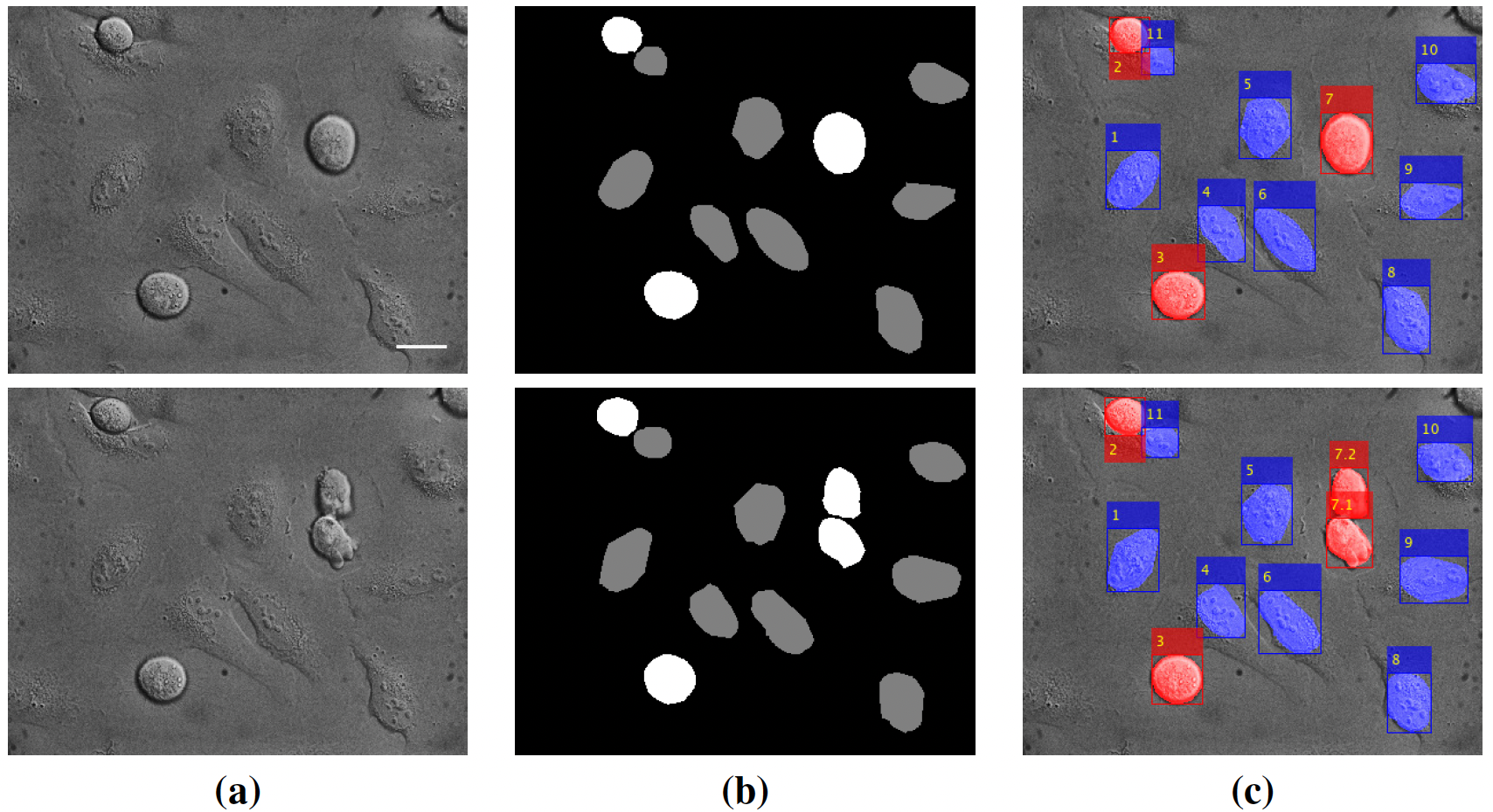 |
Label-free sequences:
Review [10] and annotated data [11],[20] for cell segmentation, event detection, and tracking for label-free microscopy imaging. |
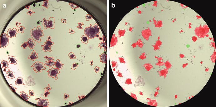 |
ESC images:
Segmentation and classification of microscopy images aimed at
identifying specific factors that control the differentiation of Embryonic Stem Cells via large-scale screenings
[3][5][7]. |
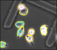 |
HeLa cell image sequences: Segmentation, tracking, and lineage of HeLa cells from phase contrast microscopy time-lapse data [4]. |
Microarray images
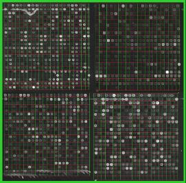 |
Automatic approach to gridding (also known as addressing or spot finding) in microarray images,
based on the Orientation Matching and the Discrete Fourier Transforms [1]. |
Magnetic Resonance images
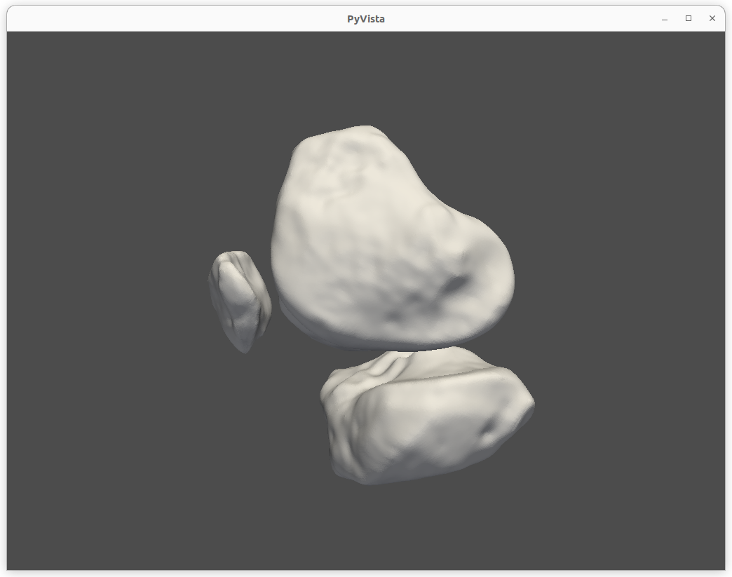 |
Segmentation and 3D reconstruction of knee bones from MRI data
[16], [17], [18], developed in the contest of the MEDIA project. |
Main collaborations:
- Computer Vision and Pattern Recognition Laboratory (CVPRLab), University of Naples Parthenope, Naples, Italy
- Dept. of Architecture, University of Florence, Florence, Italy
- Dept. of Architecture, University of Naples Federico II, Naples, Italy
- Dept. of Biology and Biotechnology, Sapienza University of Rome, Rome, Italy
- Dept. of Dermatology, Second University of Naples, Naples, Italy
- Dept. of Political Science J. Monnet, Second University of Naples, Caserta, Italy
- Esaote S.p.A., Naples, Italy
- Fondazione IDIS - Città della Scienza, Naples, Italy
- Inst. of Biostructure and Bioimaging, National Research Council, Naples, Italy
- Inst. of Genetics and Biophysics, National Research Council, Naples, Italy
- Inst. of Molecular Biology and Pathology, National Research Council, Rome, Italy
- Istituto Nazionale Tumori - IRCCS - Fondazione G. Pascale, Naples, Italy
- Stazione Zoologica Anton Dohrn, Naples, Italy
- University of Cassino and Southern Lazio, Cassino, Italy
Useful/downloadable material:
Software:
- SDI+: Matlab code implementing the algorithm for
segmentation of dermoscopic images presented in [8]
- KneeBones3Dify [18]: Python code implementing the algorithms for segmentation and 3D reconstruction of knee bones from MRI data presented in [16].
Data:
- ALFI: dataset of images and annotations for label-free microscopy presented in [11]
- KneeBones3Dify dataset [17]: dataset of MR images and annotations used for validating the knee bones segmentation and 3D reconstruction algorithms presented in [16].
Publications and Communications on Biomedical Imaging:
[20]
F. Polverino, L. Antonelli, A. Albu, A. Hada, I.A. Asteriti, F. Degrassi, L. Maddalena, M.R. Guarracino, G. Guarguaglini,
The ALFI database: images and annotations for label-free time-lapse microscopy,
Poster at
Bioinformatics and Computational Biology Conference 2024
(BBCC2024), Naples, November 27-29, 2024.
[19]
-- L. Maddalena, L. Antonelli, F. Polverino, I.A. Asteriti, F. Degrassi, G. Guarguaglini, A. Albu, A. Hada, and M.R. Guarracino,
Machine Learning and Artificial Intelligence for Cell Biology Imaging,
keynote talk at
BIOMAT 2024, Kolympari, Chania, Greece, 27/10-1/11/2024.
[18]
G. De Lucia, L. Maddalena, D. Romano, F. Gregoretti, L. Antonelli,
KneeBones3Dify 1.0 (v1.0),
Zenodo, DOI: 10.5281/zenodo.10522876, 2024.
[17]
E. Soscia, D. Romano, L. Maddalena, F. Gregoretti, G. De Lucia, L. Antonelli,
KneeBones3Dify-Annotated-Dataset v1.0.0 (V1.0.0),
Zenodo, DOI: 10.5281/zenodo.10534328, 2024.
[16]
L. Maddalena, D. Romano, F. Gregoretti, G. De Lucia, L. Antonelli, E. Soscia, G. Pontillo, C. Langella, F. Fazioli, C. Giusti, R. Varriale,
KneeBones3Dify: Open-source software for segmentation and 3D
reconstruction of knee bones from MRI data,
SoftwareX 27, 101854, ISSN: 2352-7110, DOI: 10.1016/j.softx.2024.101854, 2024.
Software available in [18].
Data available in [17].
[15]
L. Maddalena, Artificial Intelligence for Cell Biology Imaging,
invited presentation at
3D High Content Imaging for the Validation of New Anti-Cancer Molecules,
Final workshop of the Regione Lazio research project
INNOVA3DIMAGING, Rome, June 5, 2024.
See the Slides and
Workshop leaflet.
[14]
L. Maddalena, L. Antonelli (Eds),
Algorithms for Biomedical Image Analysis and Processing,
Reprint of the Special Issue, Algorithms, ISBN: 978-3-0365-9760-7, DOI: 10.3390/books978-3-0365-9761-4, 2024.
[13]
L. Antonelli, M.R. Guarracino, T. Klinsuwan, L. Maddalena,
Computational and Mathematical Methods for Biomedical Imaging,
Keynote talk at 23rd International Symposium on Mathematical and Computational Biology (BIOMAT 2023), November 2023.
[12]
L. Antonelli, L. Maddalena,
Special Issue on "Algorithms for Biomedical Image Analysis and Processing",
Algorithms 16(12), Editorial,
DOI: 10.3390/a16120544, 2023.
[11]
L. Antonelli, F. Polverino, A. Albu, A. Hada, I.A. Asteriti, F. Degrassi, G. Guarguaglini, L. Maddalena, M.R. Guarracino,
ALFI: Cell cycle phenotype annotations of label-free time-lapse imaging data from cultured human cells,
Scientific Data 10(677),
DOI: 10.1038/s41597-023-02540-1, 2023.
The ALFI dataset is available here.
[10]
L. Maddalena, L. Antonelli, A. Albu, A. Hada, M.R. Guarracino,
Artificial Intelligence for Cell Segmentation, Event Detection, and Tracking for Label-free Microscopy Imaging,
Algorithms, DOI: 10.3390/a15090313, vol. 15, no. 313, 2022.
[9] L. Maddalena and L. Antonelli (eds.),
Special Issue on Algorithms for Biomedical Image Analysis and Processing,
Algorithms, 2022.
[8] M. R. Guarracino and L. Maddalena, SDI+: a Novel Algorithm for Segmenting Dermoscopic Images, IEEE Journal of Biomedical and Health Informatics, 23(2), 481-488, 2019.
[7] L. Casalino, M. R. Guarracino, and L. Maddalena, Imaging for High-Throughput Screening of Pluripotent Stem Cells, SIAM Conference on Imaging Science - IS18, Bologna, June 2018.
[6] M. R. Guarracino and L. Maddalena, Segmenting Dermoscopic Images, arXiv:1703.03186, 2017.
[5] L. Casalino, P. DAmbra, M. R. Guarracino, A. Irpino, L. Maddalena, F. Maiorano, G. Minchiotti, E. J. Patriarca, Image Analysis and Classification for High-Throughput Screening of Embryonic Stem Cells, in V. Zazzu, M.B. Ferraro, and M.R. Guarracino (Eds.), Mathematical Models in Biology, Springer, 17-31, 2015.
[4] M. Sangiovanni, L. Maddalena, M. Guarracino, Following the Changes: HeLa cells Lineage from Phase Contrast Microscopy Time-Lapse Data in 6th International Workshop on Data Analysis Methods for Software Systems, p. 45, December 2014.
[3] Image Segmentation and Classification for High-Throughput Screening of Microscopy Imagery, Conference Horizon 2020@DIITET, CNR, May 26-27, 2014.
[2] V. Cozza, M.R. Guarracino, L. Maddalena, A. Baroni, Dynamic Clustering Detection through Multi-valued Descriptors of Dermoscopic Images, Statistics in Medicine, John Wiley & Sons, Ltd., Vol. 30, Issue 20, pagg. 25362550, DOI: 10.1002/sim.4285, 2011. [Impact Factor: 2.328]
[1] L. Maddalena, A. Petrosino, Metodi per l'Analisi di Immagini da Microarray, Tutorial Metodi e Strumenti per lanalisi dei Dati di Espressione Genica, Naples, December 2007.
Last update: January 13, 2025